
A 56-year-old patient presents with sudden blurry vision upon awakening. He has a snoring history. Why?
Analysis
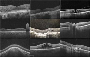
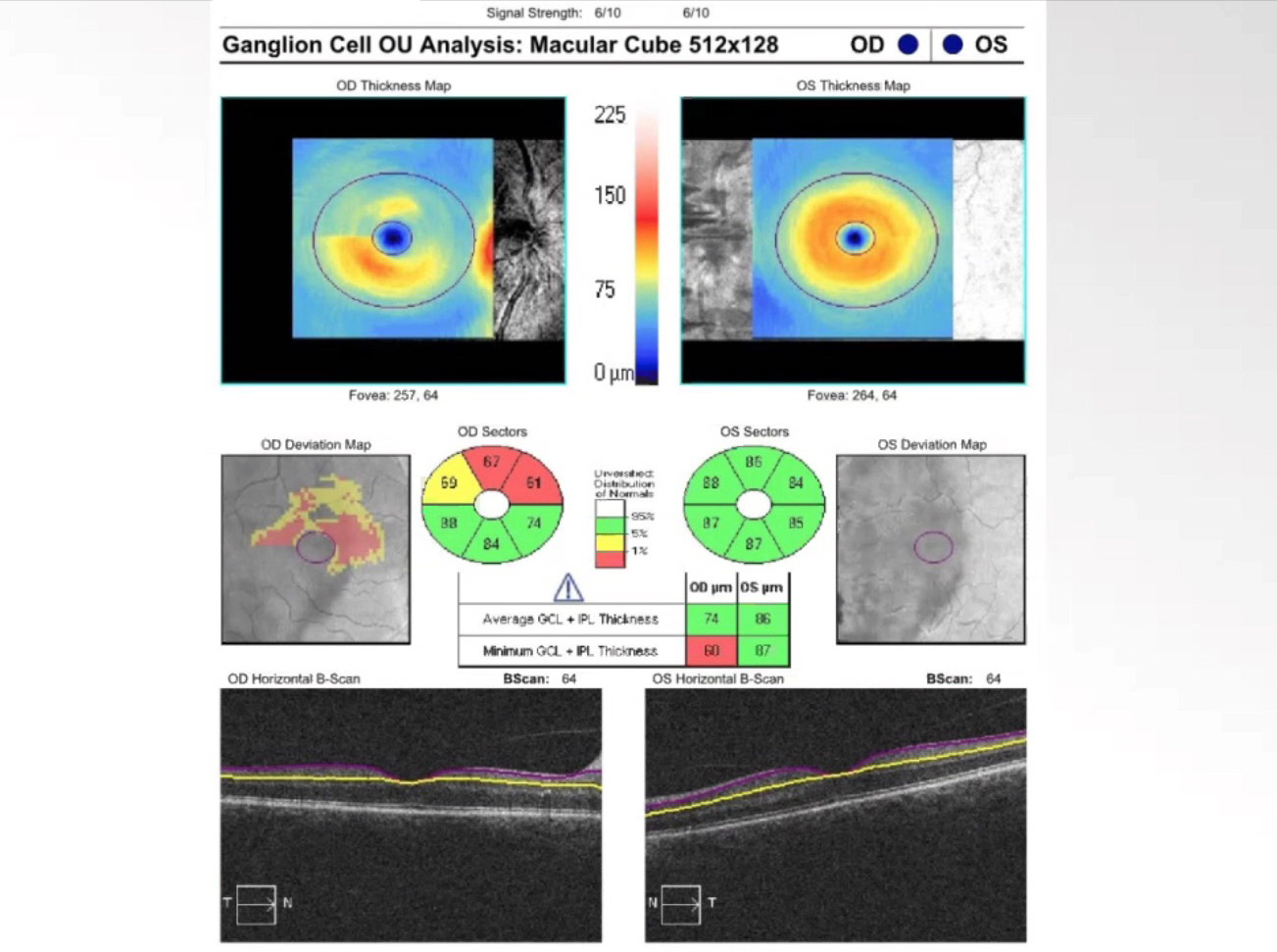
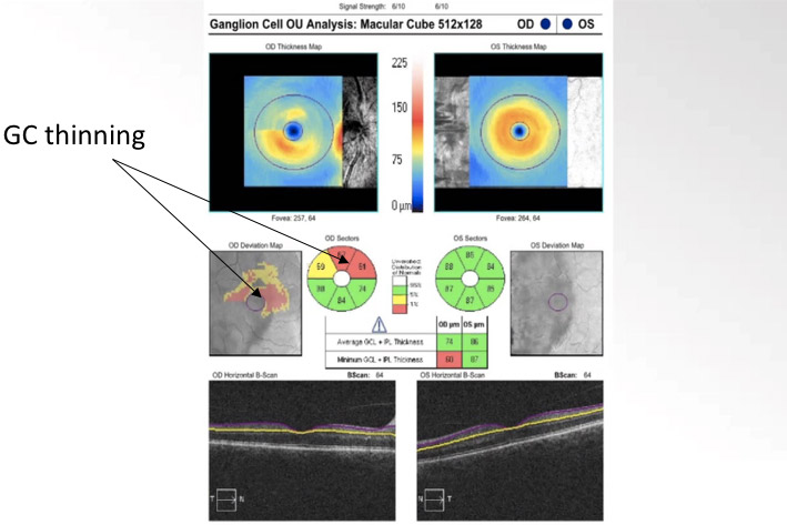
This is an OCT focusing on a ganglion cell analysis. There is evidence of nerve-fiber-layer thinning in the right eye (left image). This defect highlighted in the superior half of the macula indicates present damage to the optic nerve. The left eye (right image) is within normal limits. The OCT of the nerve also showed some swelling changes (video).
The patient was diagnosed with Non-arteritic Ischemic Optic Neuropathy (NAION). Sleep apnea is one of the known risk factors for this condition.
In the accompanying 19 min. video featuring Dr. Paul Freund, Assistant Professor at Dalhousie University in Halifax, you will learn:
- The signs on OCT ganglion cell analysis that suggest NAION
- The key questions to ask to rule out GCA
- Management of Non-arteritic Ischemic Optic Neuropathy
Neuro Coach Tip
Fifteen percent of NAION patients will develop bilateral disease within 5 years, so addressing modifiable risk factors in of critical importance.
Not receiving our tips yet? Subscribe to receive them straight to your inbox.
