Issue 6
Ganglion cell analysis on OCT
Issue 5
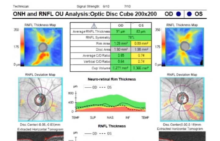
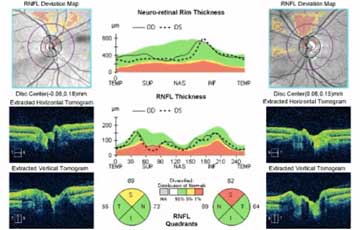
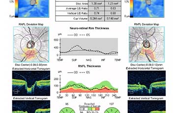
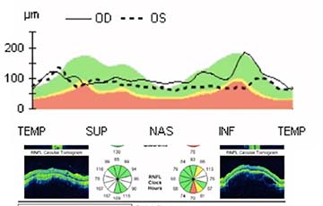
Reading an OCT in a glaucoma patient
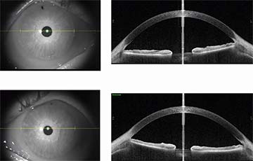
Issue 4
Is this angle open or closed?
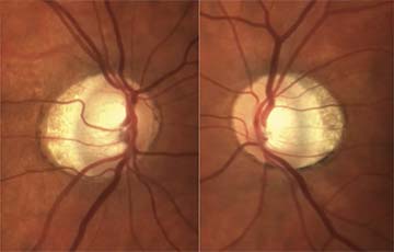
Issue 3
Optic discs in a patient with high myopia
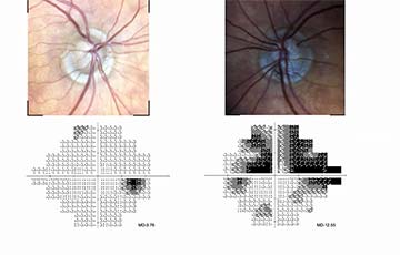
Issue 2
Abnormal visual field
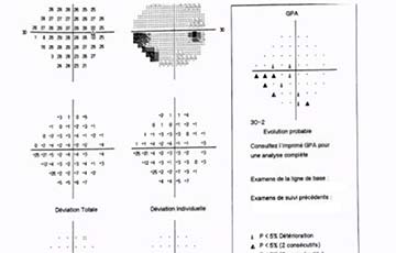
Issue 1
Is this visual field normal?
1 of 1
- 1