Enter Optocase
Login to your account or join for free now,
to access case content.
Disclosures: Dr. Sharma is the Editor-in-Chief of Optocase, and has received research grants from both Bayer and Novartis.
In this video case-based course, Dr. Sanjay Sharma will walk participants through the important aspects of several retina cases. Dr. Sharma will present and review clinical images from 15 different cases related to a variety of retinal pathologies. Key findings and diagnoses will be derived from OCTs, OCTAs, angiograms, fundus images, and other ocular scans.
Some of the topics that will be discussed include
Before moving on to the lecture, take a moment to examine the image below. An 82-year-old suddenly loses their vision. Why?
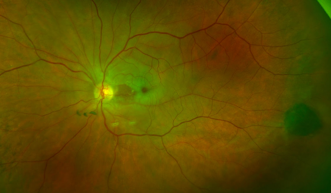
Watch the full course lecture video to explore this case further and review four important facts about retinal artery occlusion with Dr. Sharma; this is the first of 15 cases he will be investigating.
Get full access to the content of this case. Purchase this case now for $39. Plus, save when you buy 3 cases.