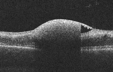

An 82-year-old was referred for a new lesion. What is your diagnosis?
Analysis
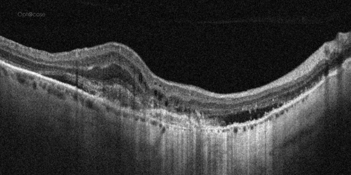
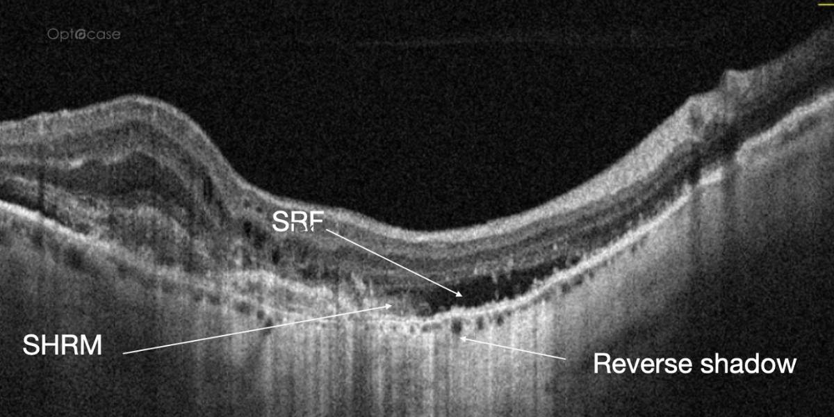
There is a significant elevation of the deep retina. There is retinal whitening which is relate to a separation of the RPE from underlying Bruch’s membrane. SRF and IRF are both seen. The OCTA showed lacy hyperreflectivity in both eyes (video). Clinical exam revealed geographic atrophy and new subretinal hemorrhage.
The patient was diagnosed with choroidal neovascular membrane associated with AMD.
In this tip's accompanying 5 min. video, we'll learn:
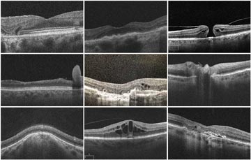
- Differential diagnoses of CNV
- Conditions that present with deep hemorrhages
- How GA appears on an OCT
OCT Tip
Always consider the diagnosis of CNV in a patient with subretinal fluid.
Not receiving our tips yet? Subscribe to receive them straight to your inbox.