Diabetic Macular Edema

    • CE credits 2 hours
    • COPE code 101107-TD / 131932
    • Available until Oct 11, 2028

Introduction

Case

A 43-year-old male presents to your clinic for his annual diabetic eye exam. When he was last seen one year ago, best-corrected visual acuity was 20/20 OU with scattered microaneurysms in both eyes. On further questioning, he notes decreased vision in the left eye over the past six months and inquires about “stronger glasses”. His sugar control has been moderate with a recent hemoglobin A1c of 8.5%.

On exam, best-corrected visual acuity is 20/20-2 in the right eye vs. 20/50 in the left eye. Intraocular pressures and pupillary responses are normal, and no evidence of significant corneal or lens opacity is seen. Fundus exam of the left eye reveals the following:
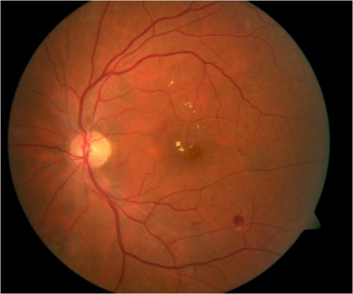
A 43-year-old male presents to your clinic for his annual diabetic eye exam.

Question

Which of the following investigations would be most useful to identify the source of the condition seen that is most likely the cause of decreased vision?

  • Optical coherence tomography

    Optical coherence tomography (OCT) is an excellent test to quantify the amount of retinal edema, but is not useful in determining the source of leakage.

  • Amsler grid

    Amsler grid would help identify the size of the patient’s central scotoma/metamorphopsia, but would not identify the source of leakage.

  • Intravenous fluorescein angiography
    CORRECT

    Intravenous fluorescein angiography (IVFA) is a critical test to determine the source of leakage with diabetic macular edema, which has important implications for macular laser photocoagulation.

  • Indocyanine green angiography

    ICG angiography is an alternative to IVFA that is excellent at studying choroidal circulation, but would typically not be used prior to IVFA.

Learning Objectives

  • To review the pathophysiology, diagnosis, and treatment options for diabetic macular edema
  • To review the safety and efficacy of anti-VEGF compounds in diabetic patients
  • To review the impact of anti-VEGF compounds on microaneurysms

Introduction

Diabetic macular edema (DME) is a frequent and important cause of vision loss in diabetic patients. It is the predominant cause of vision loss in patients with non-proliferative diabetic retinopathy (NPDR) as well as diabetic retinopathy as a whole.1 In addition, it is also the most common cause of vision loss in the working age population in developed countries,2 affecting 3-6% of diabetics over the age of 18.2 It is more common in type II compared with type I diabetics.3

The presence of DME is strongly correlated with duration of diabetes and glycemic control. Its prevalence increases from 5% of individuals at 5 years after diagnosis to 15% at 15 years.4 It affects roughly 3% of individuals with mild NPDR, 38% with moderate-severe NPDR and 71% with proliferative disease. Roughly 75,000 new cases of DME are diagnosed in the USA each year.5

This module will focus on the pathophysiology, diagnosis, and management of DME, with a special focus on the use of anti-VEGF agents.

Access Full Case Content

Get full access to the content of this case. Purchase this case now for $29. Plus, save when you buy 3 cases.

See all cases