Enter Optocase
Login to your account or join for free now,
to access case content.


In this video case-based course, Dr. Sanjay Sharma, Dr. Christopher Rapuano, and Dr. Melissa Barnett will walk participants through the important aspects of several cornea-related clinical cases. Test your knowledge with this quiz-style presentation where the lecturers will show a clinical image, allow time for the viewer to attempt a diagnosis, and then review the correct answer.
In this video, the lecturers will review:
Before moving on to the lecture, take a moment to examine the image below. An 82-year-old presents with a chronic red eye. What corneal pathology is noted?
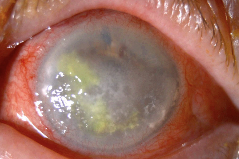
Herpes simplex is a viral infection in which there is activation of the virus in the sensory nerve fibers. Classically, epithelial involvement is seen with a dendritic pattern of staining. This is not present here.
Band keratopathy is the correct answer. In this condition, calcium deposits in Bowman's layer, the epithelial basement membrane and the anterior stroma. This is a form of dystrophic calcification as calcium is deposited in tissue that is degenerated from a variety of causes including systemic or local inflammation, infection, medication or injury.
Munson's sign is seen in keratoconus. Here when a patient looks down, an abnormally shaped cornea causes a v-shaped distortion of the lower lid.
Mooran's ulcer is a peripheral ulcer; the pathology here is central in nature. Additionally, the image is not consistent with an ulcer.
Disclosures: Dr. Sharma is the Editor-in-Chief of Optocase, and has received research grants from both Bayer and Novartis.
Disclosures: Dr. Barnett has received honorariums for various roles from: ABB, Acculens, Allergan, Azura, Bausch + Lomb, BCLA, Bruder, CooperVision, Dompé, EveryDay Contacts, Gas Permeable Lens Institute, JJVC Vistakon, Lentechs, Mojo Vision, Novartis, Ocusoft, Prasis, Oyster Point, Percept, RVL Pharmaceuticals, Sight Sciences, Science Based Health, Scleral Lens Education Society, Sjogren's Syndrom Foundation, STAPLE program, Sun Pharma, Tarsus, Tarsus, Visus Therapeutics, Vyluma.
Disclosures: Dr. Rapuano has been a consultant for the following: Avellino.
Get full access to the content of this case. Purchase this case now for $39. Plus, save when you buy 3 cases.