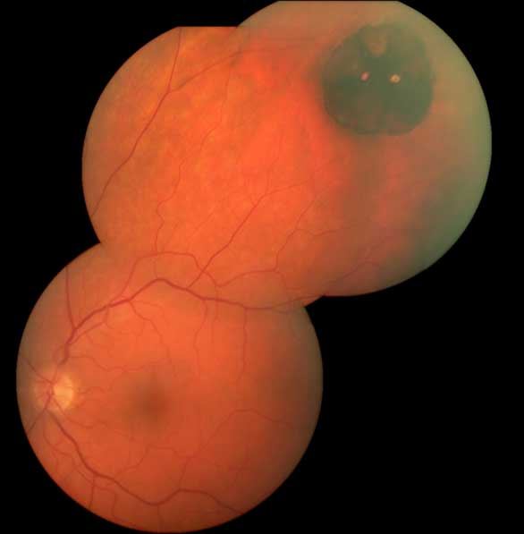
A 40-year-old female presents to your office for a routine eye exam. She has no specific concerns. She has not seen an optometrist in over 20 years, and has no history of any eye problems. She is healthy apart from well-controlled hypertension and migraines. On dilated exam, a large pigmented lesion is seen in the superotemporal retina.
Introduction
Objectives
- To review the demographics, pathophysiology, and characteristics of congenital hypertrophy of the retinal pigment epithelium (CHRPE)
- To review the association between CHRPE and colon cancer
- To review the features and purpose of a Cochrane Review
Case
Quick Question
Examine the following image of the above patient. What is the most likely diagnosis?
-
Choroidal melanoma
Choroidal melanoma should be considered in the differential diagnosis of any pigmented fundus lesion. Choroidal melanoma is less likely in this case due to the absence of drusen, orange pigment, or elevation of the lesion.
-
Choroidal nevus
Choroidal nevus is a common cause of pigmented fundus lesions. However, the appearance in this example is more in keeping with CHRPE.
-
Congenital hypertrophy of the retinal pigment epithelium
Correct -
RPE adenocarcinoma
Although extremely rarely RPE adenocarcinoma may arise from a CHRPE lesion, there are no signs to suggest this is the case in this example. Specifically, the lesion here is not raised and there does not appear to be abnormal vasculature supplying the lesion.
Introduction
Congenital hypertrophy of the retinal pigment epithelium (CHRPE) is a benign lesion of the fundus that most often appears as a flat, pigmented spot.1 Its prevalence is highly variable and difficult to determine- studies quote a highly variable prevalence ranging from 0.3-40% of the population.2-4 It is more common in females by a ratio of 2:1.5
CHRPE can be broken down into three subtypes which include solitary CHRPE (which is most often referred to with the term “CHRPE”), grouped CHRPE (“bear tracks”), and multiple CHRPE. The latter 2 subtypes are often considered distinct entities, and this module will focus primarily on the solitary type. Typically, CHRPE has no clinical significance, however the recent development of multiple CHRPE is often associated with the cancer syndrome familial adenomatous polyposis (FAP).1
Access Full Case Content
Get full access to the content of this case. Purchase this case now for $29. Plus, save when you buy 3 cases.