Retinal Vein Occlusion

    • CE credits 3 hours
    • COPE code 86634-SD / 127968
    • Available until Mar 1, 2027

Introduction

Learning Objectives

  • To review the epidemiology, pathophysiology, and findings of retinal vein occlusion
  • To review treatment options for retinal vein occlusion
  • To review results of the CRYSTAL study

Case

A 64-year-old male is referred to your office on an urgent basis by their family doctor for a possible retinal detachment. On history, the patient complains of awakening with a “film” over his inferior visual field of the right eye. He has complained of occasional photopsia but no floaters. The patient's past ocular history is unremarkable. Past medical history is significant for hypercholesterolemia and well-controlled hypertension.

On exam, best-corrected visual acuity is 20/25 OD vs. 20/20 OS. There is no relative afferent pupillary defect and IOPs are within normal limits. The patient can count fingers in all 4 quadrants with both eyes.

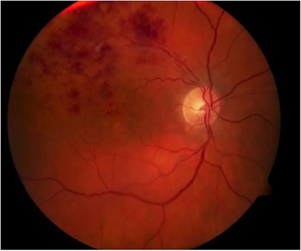
Anterior segment exam is unremarkable. Posterior segment exam of the right eye is shown below:

Quick Question

Apart from increasing age, what is the most important/common risk factor for the patient's condition?

  • Hypercholesterolemia

    Hypercholesterolemia is an important risk factor for RVO, especially in younger patients. However, hypertension is still a more important risk factor for RVO, especially in a patient over the age of 50.

  • Obesity

    High body mass index (BMI) is a risk factor for a multitude of vascular events both within and outside the eye, but is less important than hypertension as a risk factor for BRVO.

  • Hypertension
    CORRECT

    The patient has a branch-retinal vein occlusion (BRVO), of which hypertension is the most important and common risk factor.

  • Glaucoma

    Glaucoma is a risk factor for both BRVO and CRVO, however hypertension is by far the most common risk factor for BRVO.

Case

Retinal vein occlusion (RVO) is a common, sight-threatening retinal disorder caused by blockage or thrombosis within the retinal venous system.1 It is second only behind diabetic retinopathy as the most common form of retinal vascular disease,2 and is estimated to affect 16.4 million adults worldwide.3

The overall prevalence of RVO is approximately 0.1-0.5% in middle to older aged groups,4, 5 with a prevalence of up to 4.6% in individuals over the age of 80.6 The 4 year incidence of RVO is estimated at 5 per 1000 individuals aged 65 and older.7 There is no racial or gender predilection.8

Typically RVO is classified by the location of occlusion into either a central retinal vein occlusion (CRVO), branch retinal vein occlusion (BRVO), or hemiretinal vein occlusion (HRVO). Although BRVO is roughly 3-4x more common than CRVO, CRVO affects anatomy and visual function more severely than BRVO.9 In fact, the dramatic appearance of CRVO is often termed “blood and thunder” and the entity itself called a “stroke of the eye” for its often sudden and severe nature.

Vision loss with RVO depends on the location and severity of venous occlusion. Macular edema and ischemic sequelae are common causes of vision loss. Traditionally, treatment options of RVO have been limited but newer treatment options such as anti-VEGF agents have significantly changed the outlook of patients with RVO.

Access Full Case Content

Get full access to the content of this case. Purchase this case now for $39. Plus, save when you buy 3 cases.

See all cases