Enter Optocase
Login to your account or join for free now,
to access case content.
Disclosures: Dr. Singh has the following financial relationships to disclose: Isoaid (consultant), Immunocore (speaker bureau), Auro (consultant). All relevant relationships have been mitigated.
In this video case-based course, Dr. Arun Singh will be teaching on the treatment for retinoblastoma with a focus on current management practices involving chemotherapy routes. The course will also review the global aspects of retinoblastoma including the burden of the disease, challenges to treatment and possible solutions. Dr. Singh will then go over the evaluation and staging for retinoblastoma, followed by the methods of treatment. The course will conclude with three illustrative cases.
Before moving on to the lecture, take a moment to examine the image below. A 3-year-old presents with an eye turning in. What is your concern?
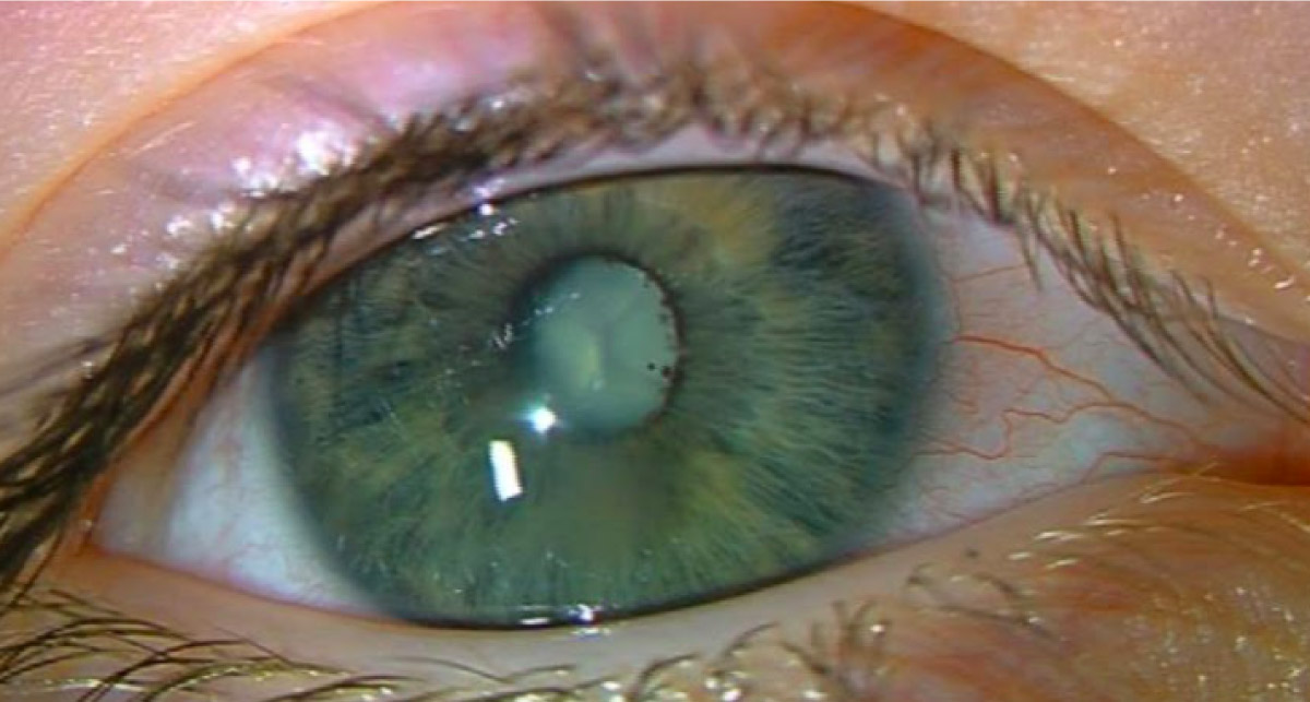
Granular dystrophy is an inherited condition in which there are discrete areas of white lesions in the central cornea. Corneal lesions are not seen here.
Nasolacrimal duct obstruction presents with redness and swelling of the medial canthal area as there is infection of the lacrimal sac. This is not seen here.
Leukocoria, the presence of a white pupil, is seen here. In addition, there is the presence of pigment on the surface of the lens, suggestive of posterior synechiae, indicating inflammation. This patient was diagnosed with retinoblastoma.
A corneal ulcer can occur from a variety of reasons including infections (including bacterial and parasitic), inflammations and trauma. There is no corneal ulcer present here as the epithelium is intact.
Get full access to the content of this case. Purchase this case now for $39. Plus, save when you buy 3 cases.