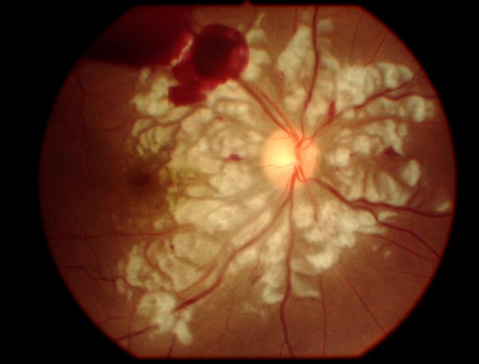
A 25-year-old woman complained of sudden loss of vision in her right eye immediately following a car accident. She had fractured both femurs and numerous ribs, but had no major facial trauma. Her vision in the affected eye was reduced to the ability to only count fingers.
Introduction
Case
Question
The photograph is consistent with which of the following conditions? Please choose between one of the following four answers. One is correct, the others are not.
-
Retinal artery occlusion
Patients with retinal artery occlusion present with sudden painless visual loss. Clinical findings include confluent cloudy swelling of the retinal nerve fibre layer which classically presents with “cloudy swelling” with the presence of a “cherry-red” spot. While there are elements of cloudy swelling in this image, they are much more segmented and include a clear zone on either side of the retinal vasculature. Additionally, a cherry-red spot, which is caused by the normal choroidal/RPE tissue being contrasted by the adjacent swollen NFL is not seen here.
-
Purtscher's retinopathy
CORRECT -
Retinal vein occlusion
Patients with retinal vein occlusion present with painless visual loss. The classical retinal picture of central retinal vein occlusion, known as “blood and thunder”, is the presence of flame-shaped, nerve-fibre layer hemorrhages. Numerous areas of retinal whitening, if present in retinal vein occlusion, are usually associated with retinal hemorrhages.
-
Myelinated nerve fibres
Myelinated nerve fibres appear as areas of retinal whitening that appear “feathery” in nature. These fibres are much more linear that the retinal swelling that is seen in retinal artery occlusion or Purtscher's retinopathy. Myelin, typically, is not located in the retina. These lesions are not of pathological significance.
Introduction
Ocular trauma can be associated with several findings including globe rupture, hyphema, angle recession, traumatic cataract in the anterior segment; retinal findings, including retinal tear and detachment, retinal “bruising” (called commotio retina and representing photoreceptor outer segment disruption), retinal dialysis (disinsertion of peripheral retina from the underlying choroid), and intraretinal or vitreous hemorrhage can also occur. Trauma distant from the eye, including chest and long-bone fracture, can also be associated with changes in the retina.