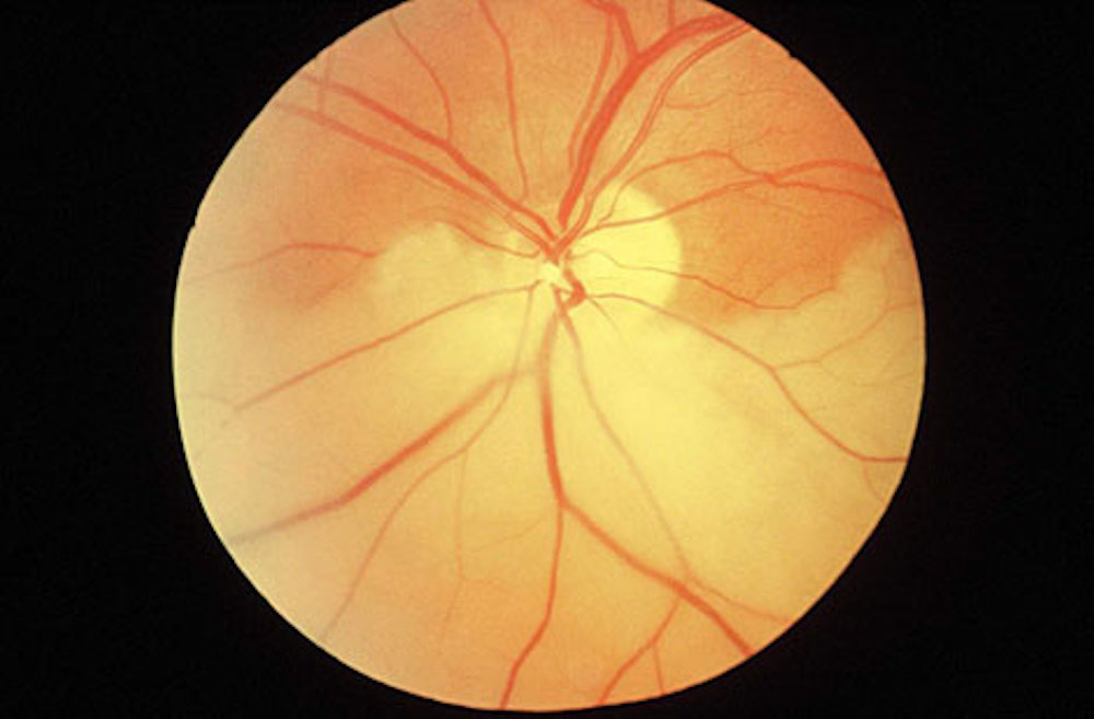
A 76-year-old female presents with a 24 hour history of decreased vision in her left eye. She says the vision loss occurred suddenly, and has not had any other symptoms such as flashes, floaters, or pain in the eye. Her ocular history is significant for cataract surgery in both eyes over ten years ago, while her medical history is significant for longstanding hypertension and dyslipidemia. On exam, visual acuities are 20/25 in the right eye and 20/40 in the left eye. Intraocular pressure and anterior segment exam are normal in both eyes. The fundus appearance of the left eye is shown below.
Introduction
Learning Objectives
- To review the epidemiology, pathophysiology and presentation of retinal artery occlusion
- To review evidence behind treatment options for retinal artery occlusion
- To review the concept of disease prevention in epidemiology
Case
Quick Question
What is the most likely diagnosis?
-
Branch retinal artery occlusion
Correct -
Central retinal artery occlusion
In the fundus photograph one can clearly see that the blockage is distal to the bifurcation of the central retinal artery. A central retinal artery occlusion would typically present with a worse visual acuity than the case in question.
-
Branch retinal vein occlusion
The fundus appearance is consistent with an arterial rather than venous occlusive pathophysiology, the latter of which classically appears as “blood and thunder.”
-
Central retinal vein occlusion
The fundus appearance is consistent with an arterial rather than venous occlusive pathophysiology, the latter of which classically appears as “blood and thunder.”
Introduction
Retinal artery occlusion is an important cause of vision loss in the elderly, and is caused by any event causing significant blockage of a retinal artery. Such a blockage is termed either a central retinal artery occlusion (CRAO) or branch retinal artery occlusion (BRAO) depending on whether the occlusion occurs before or after the bifurcation of the retinal artery, respectively.
CRAOs occur at an incidence of approximately 0.85 cases per 100,000 per year,1 with BRAOs being less common. Of all retinal artery occlusions, CRAOs account for 58% of cases, BRAOs for 38% and cilioretinal artery occlusions for the remaining 5%.2 There is a slight male predominance.
Access Full Case Content
Get full access to the content of this case. Purchase this case now for $29. Plus, save when you buy 3 cases.