
Glaucoma Coach #8 was originally published on October 10th, 2020.
This 76-year-old female presents with a new blurry spot in her left eye. What do you think is going on?
Analysis
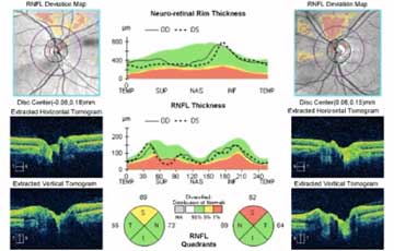
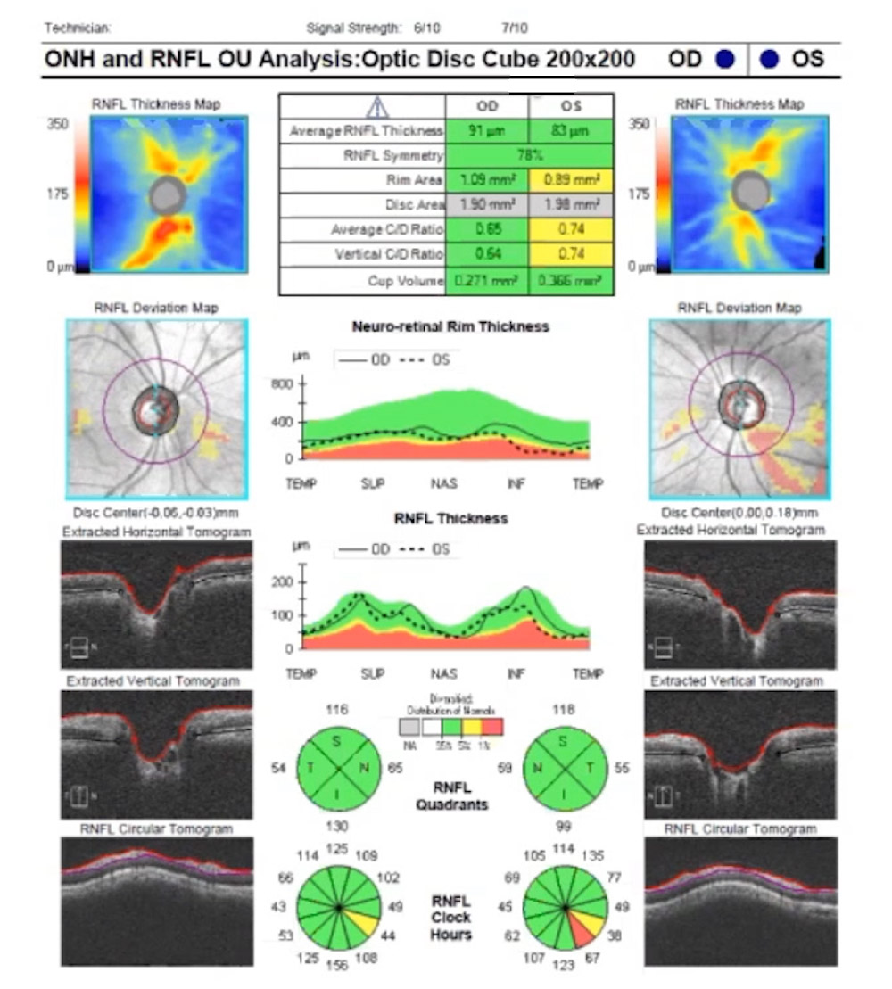
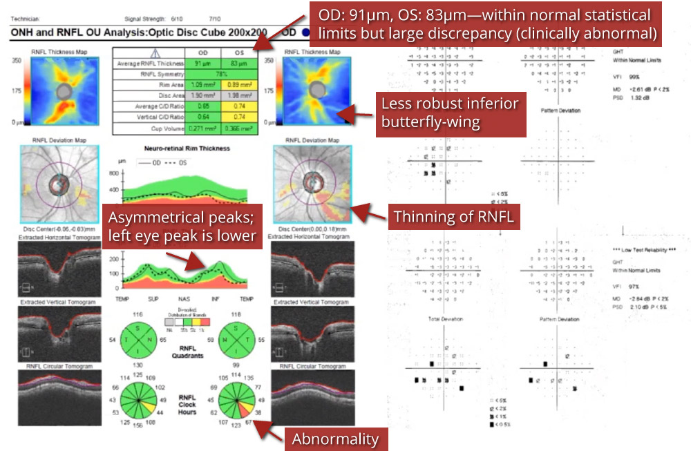
Here, we see a statistically normal but clinically abnormal retinal nerve fibre layer (RNFL) analysis. Although most of the RNFL analysis is within normal limits, there are significant discrepancies between the right and left eyes.
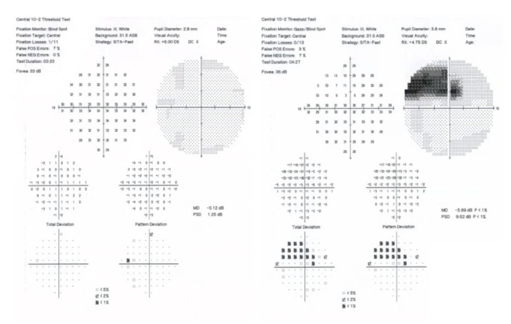
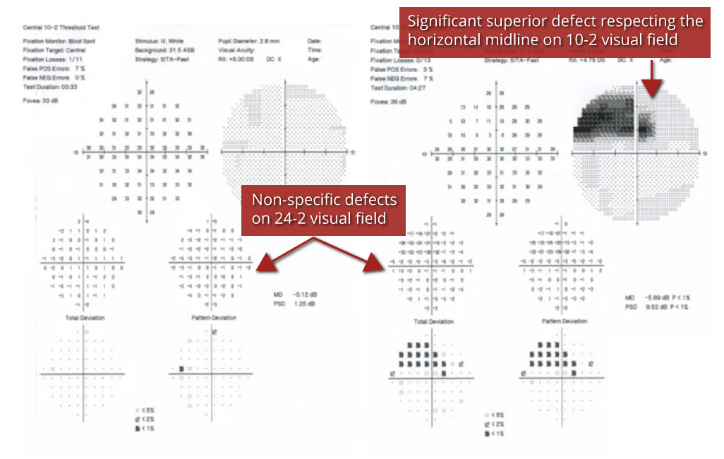
This patient's 24-2 visual field test was inconclusive, so a 10-2 visual field test was performed, revealing a significant superior defect in the left eye respecting the horizontal midline—a classic finding in glaucoma.
In this tip's accompanying 8 min. video, featuring Dr. Hady Saheb, Glaucoma Fellowship Director at McGill University in Montreal, we will review:
- The importance of evaluating discrepancies in statistically normal RNFL analysis
- The diagnostic value of 10-2 visual field testing
- Caveats of ganglion cell analysis
Glaucoma Coach Tip
Order a 10-2 visual field when a 24-2 visual field is inconclusive or inconsistent with your patient's symptomology; assess statistically normal RNFL values when the index of suspicion for glaucoma is high.
Not receiving our tips yet? Subscribe to receive them straight to your inbox.