
Glaucoma Coach #7 was originally published on September 1st, 2020.
This patient was referred as a glaucoma suspect. Why?
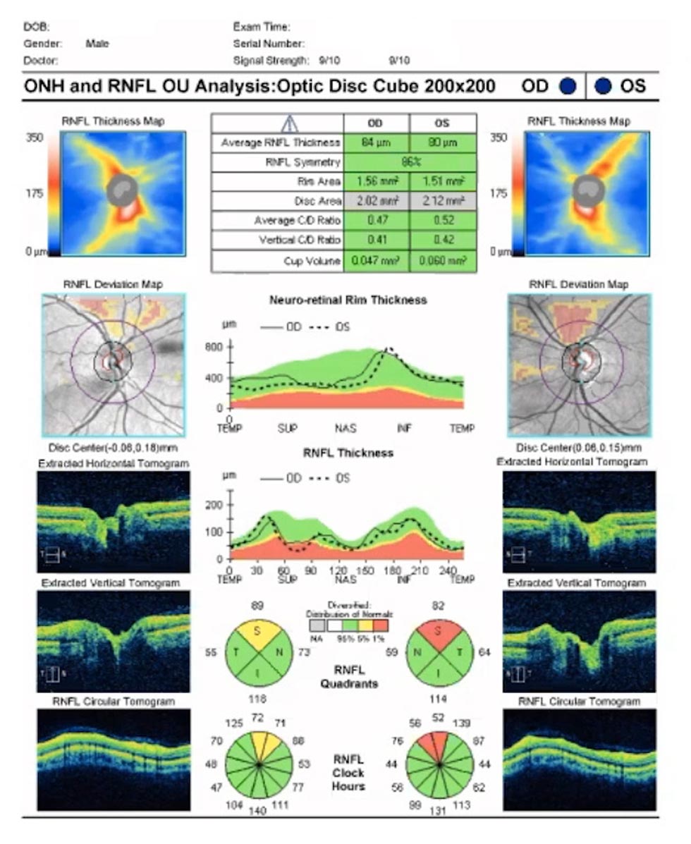
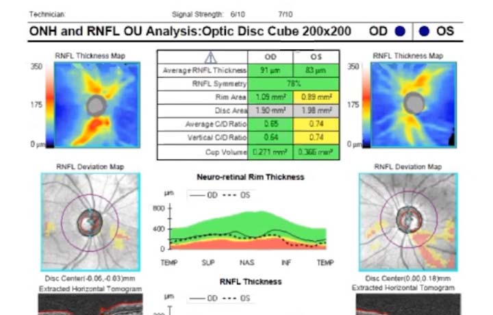
Analysis
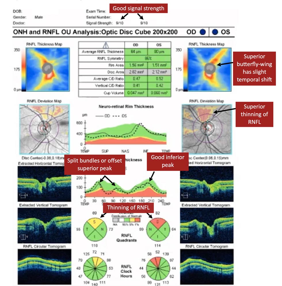
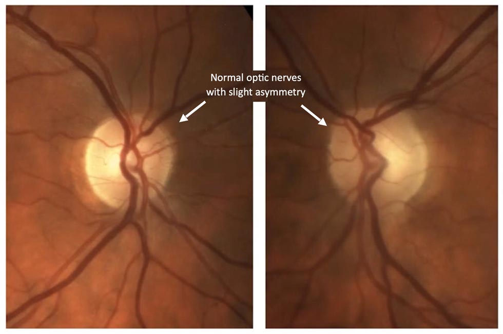
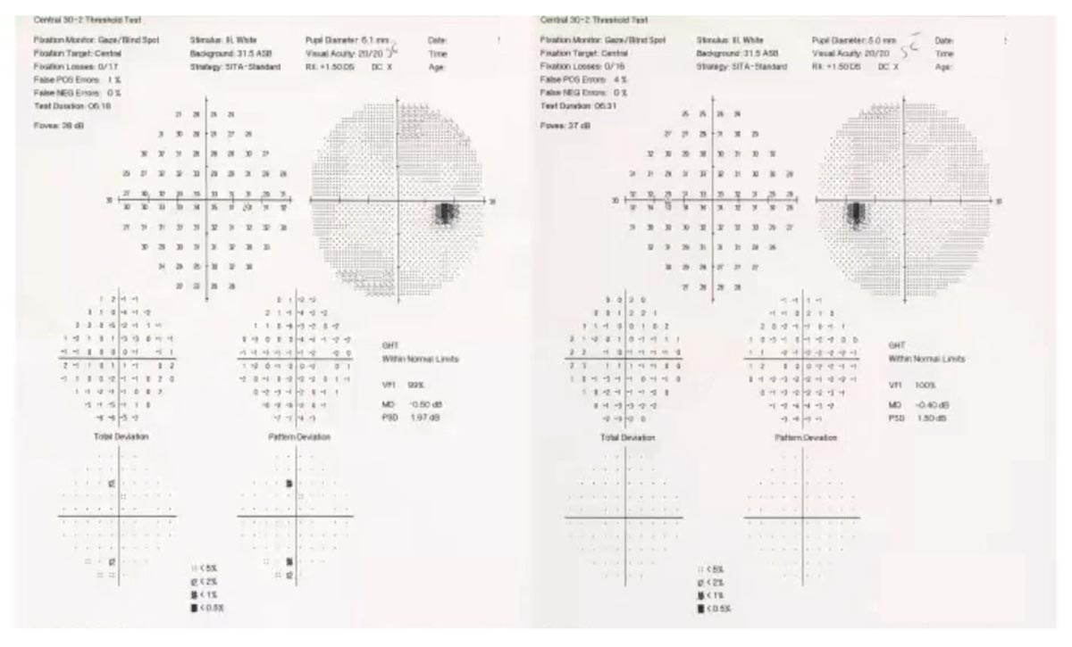
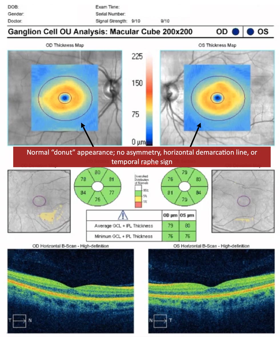
Here, we see a patient with statistical abnormality of the retinal nerve fibre layer (RNFL) on OCT but normal optic nerves, visual fields, and ganglion cell analysis. The statistical abnormality arose due to split bundles or off-setting of the superior peak on RNFL thickness analysis when compared to age-matched controls. This resulted in a false positive for glaucoma. Split bundles are a common finding in myopes.
In the accompanying 7 min. video featuring Dr. Hady Saheb, Glaucoma Fellowship Director at McGill University in Montreal, you will learn:
- The importance of assessing RNFL thickness profiles
- How to recognize a false positive glaucoma suspect on RNFL analysis
- That myopes are susceptible to split bundles on RNFL analysis
Glaucoma Coach Tip
Statistical abnormalities do not necessarily equate to clinical abnormalities (not all “red” means abnormal); split bundles can be a normal finding in myopes.
Not receiving our tips yet? Subscribe to receive them straight to your inbox.