
A young patient presented with a central elevation. What is it?
Analysis
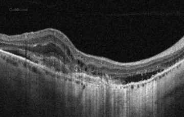
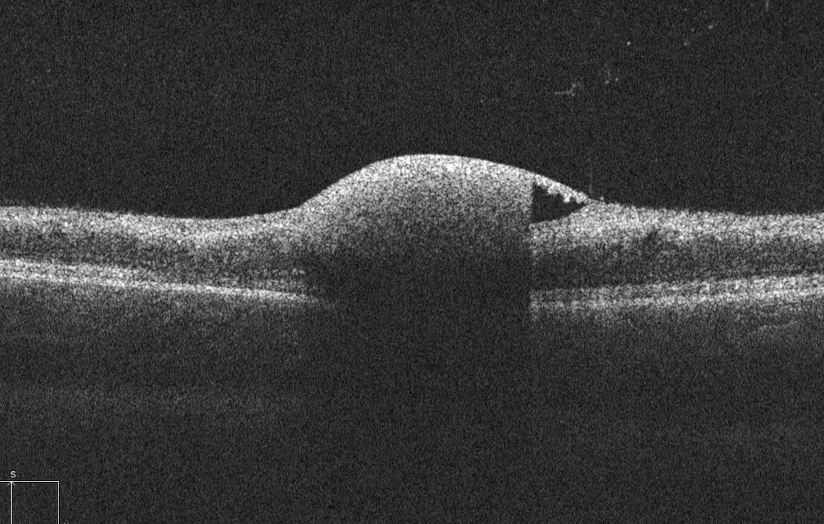
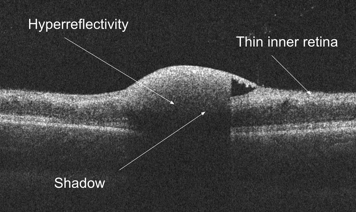
In this vertical cut, there is a clear elevation of the inner retina. The lesion is hyper reflective anteriorly. The hyperreflectivity is associated with a deeper shadow. The location of the hyperreflectivity is in the subhyaloid space. The retina is generally thinned.
This patient was noted to have a previous central retinal artery occlusion (CRAO) and numerous preretinal hemorrhages (see video).
In this tip's accompanying 13 min. video, we'll review:
- Common location of retinal hemorrhage
- Causes of retinal artery occlusion in patients younger than 30
- Differential diagnosis of superficial retinal hemorrhages
OCT Tip
In a patient with poor vision and inner retinal thinning, consider the diagnosis of previous retinal artery occlusion.
Not receiving our tips yet? Subscribe to receive them straight to your inbox.