
OCT Tip 178 was originally published on May 19th, 2020.
A 70-year-old presents with blurred vision and headache. Why?
Analysis
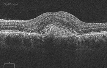
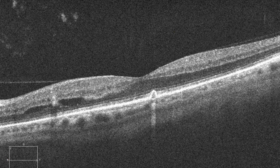
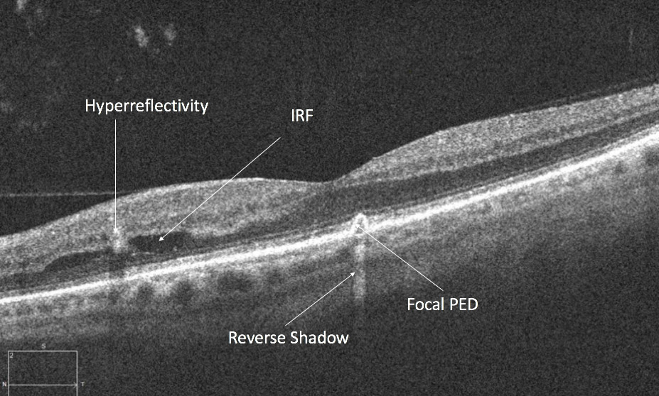
In this OCT, there is evidence of some cystic fluid located in the outer nuclear layer. In addition, there is a hyperreflective lesion in this area (left of image) and some adjacent inner nuclear layer hyporeflectivity (hemorrhage and fluid). There is also a small focal pigment epithelial detachment which is associated with some hyperreflectivity. The patient was also noted to have disc swelling and a serous detachment in the other eye (video). Their blood systolic and diastolic pressure was significantly elevated.
They were diagnosed with malignant hypertension.
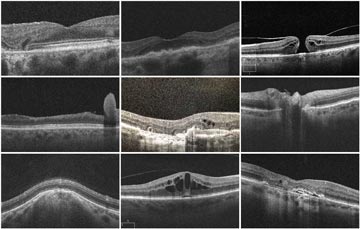
In this tip's accompanying 19 min. video, Dr. Sharma takes you through additional diagnostic imagery as well as:
- Review the inner and outer blood retinal blood barrier
- The risk of stroke with hypertensive retinopathy
- Review the OCTA findings with hypertensive choroidopathy
OCT Tip
In a patient with disc swelling and serous retinal detachment, ruling out hypertension is a must.
Not receiving our tips yet? Subscribe to receive them straight to your inbox.