-
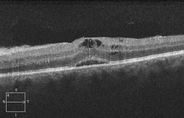
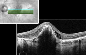
 #52. Deep Retinal Fluid: Is it RVO?Archived
 -
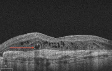
 #51. Reflective spots in outer retinaArchived
 -
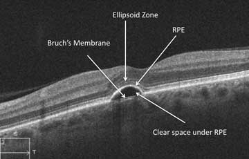
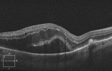
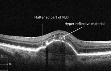
 #50. A pigment epithelial detachment (PED) with homogenous materialArchived
 -
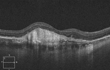
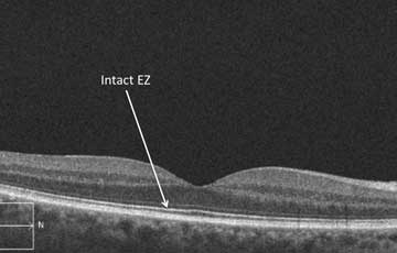
 #49. The 3rd Deep LineArchived
 -
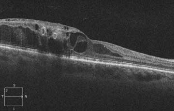
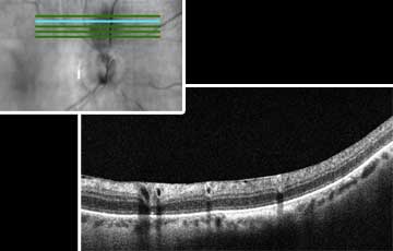
 #48. Look at the choriocapillaris in choroidal elevationsArchived
 -
 #47. Thin central retina with yellow flecksArchived
 -
 #46. A bright line on the surface of the retinaArchived
 -
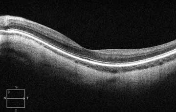
 #45. Look at the choroid on each OCT that you interpretArchived
 -
 #44. Hyper-reflectivity on surface of retinaArchived
 -
 #43. Which eye in the OCT?Archived
 -
 #42. Hyperreflectivity in the vitreousArchived
 -
 #41. Superficial hyperreflectivity with little deep retinal detailArchived
 -
 #40. Bright donuts in inner retinaArchived
 -
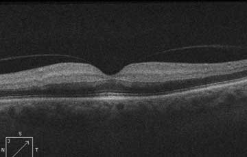
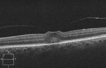 #39. A flat foveal surfaceArchived
 -
 #38. A series of hyperreflectivity lesions in superficial retinaArchived
 -
 #37. A pigmented lesion near the optic discArchived
 -
 #36. Lamellar bands below the retinal pigment epithelium (RPE)Archived
 -
 #35. A yellow lesion in the deep retinaArchived
 -
 #34. Hole in optic nerveArchived
 -
 #33. Outer retinal cysts with crystalsArchived
 -
 #32. Intraretinal cysts following cataract surgeryArchived
 -
 #31. Central cysts in the retinaArchived
 -
 #30. Dark intraretinal circles on OCTArchived
 -
 #29. Hyperreflectivity with posterior vitreous detachment (PVD)Archived
 -
 #28. Absence of retinal tissue in foveaArchived
 -
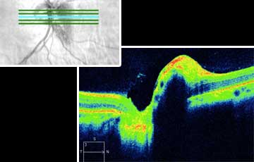
 #27. A choroidal elevationArchived
 -
 #26. Hyperreflective lesions in an overweight middle aged personArchived
 -
 #25. SRF with PEDArchived
 -
 #24. Choroidal neovascular membrane (CNV) with a thin choroidArchived
 -
 #23. Hyperintense OCT material is not always exudate or hemorrhageArchived
 -
 #22. Cystic pigment epithelial detachment (PED)Archived
 -
 #21. A hyperreflective lesion in the superficial retinaArchived
 -
 #20. Irregular pigment epithelial detachment (PED)Archived
 -
 #19. On every OCT look at the choriocapillarisArchived
 -
 #18. Always look at the choroid in all patients with subretinal fluid (SRF)Archived
 -
 #17. Numerous white spots in the retinaArchived
 -
 #16. Don't assume that subretinal fluid (SRF) equals wet age-related macular degeneration (AMD)Archived
 -
 #15. Thickened inner retinaArchived
 -
 #14. Fluid or normal?Archived
 -
 #13. Is hyperreflectivity in retina always abnormal?Archived
 -
 #12. Pigment epithelial detachment (PED) or a rip?Archived
 -
 #11. Deep circular rims of hyperreflectivityArchived
 -
 #10. Where does fluid usually accumulate?Archived
 -
 #9. Large white mound under retinaArchived
 -
 #8. Don't just look at central macular thickness (CMT)!Archived
 -
 #7. A bright deep band on OCTArchived
 -
 #6. What is subretinal hyper-reflective material (SHRM)Archived
 -
 #5. A hyperreflective spot in the retinaArchived
 -
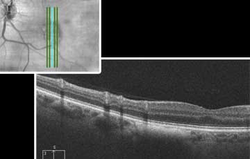
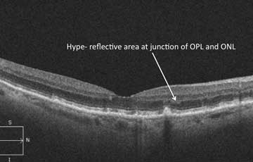 #4. Brightness in the outer plexiform layer (OPL)Archived
 -
 #3. How to predict vision from an OCTArchived