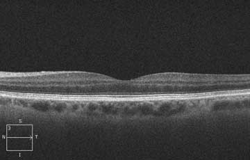
Issue 49
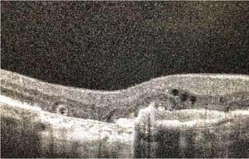
The 3rd Deep Line
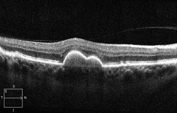
Issue 50
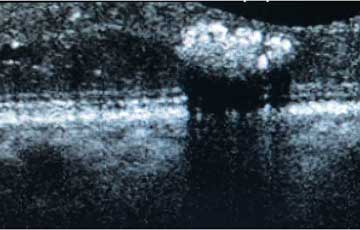
A pigment epithelial detachment (PED) with homogenous material
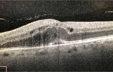
Issue 51
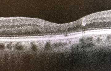
Reflective spots in outer retina
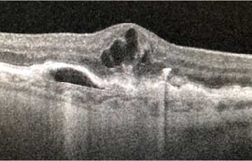
Issue 52
Deep Retinal Fluid: Is it RVO?
Issue 53
Why sudden visual loss after eye injection?
Issue 54
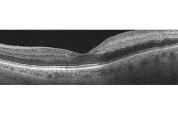
A line in the middle of the retina
Issue 55
Thin outer retina
Issue 56
What is this white lesion?
Issue 57
A 47-year-old with butterfly-shaped lesions
Issue 58
Hyperreflectivity anterior to the retina
Issue 59
Cystic spaces in 2 layers of the retina
Issue 60
Line on Retinal Surface