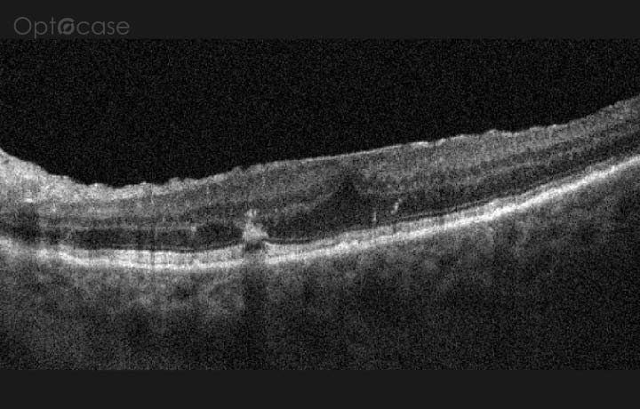
Issue 300
46-year-old with new blurring
Issue 111
88-year-old with sudden visual loss
Issue 57
A 47-year-old with butterfly-shaped lesions
Issue 7
A bright deep band on OCT
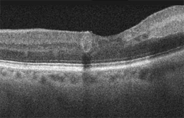
Issue 46
A bright line on the surface of the retina
Issue 27
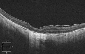
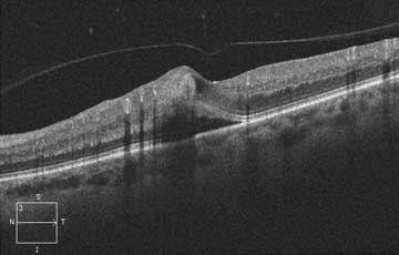
A choroidal elevation
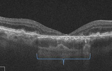
Issue 204
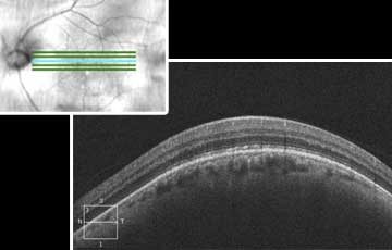
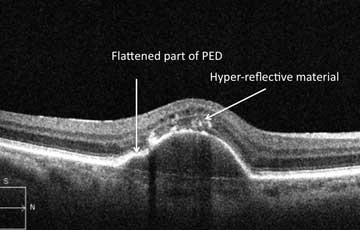
A Deep Elevation
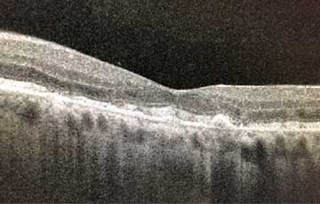
Issue 67
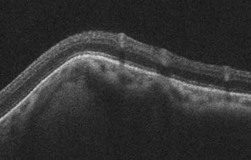
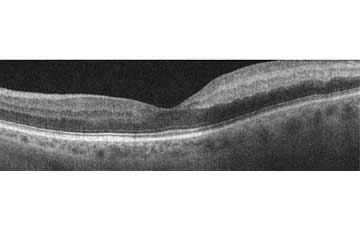
A Deep Retinal Elevation
Issue 39
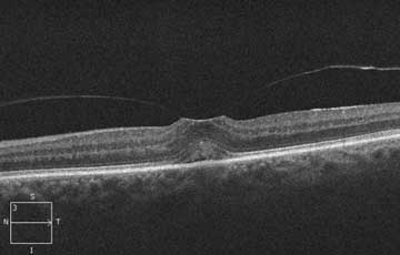
A flat foveal surface
Issue 21
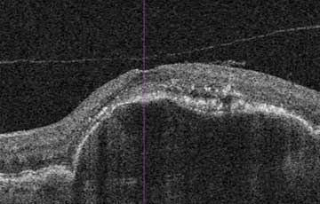
A hyperreflective lesion in the superficial retina
Issue 5
A hyperreflective spot in the retina
Issue 54
A line in the middle of the retina