Diagnostic Imagery Atlas Join the OCT Academy program to access the atlas
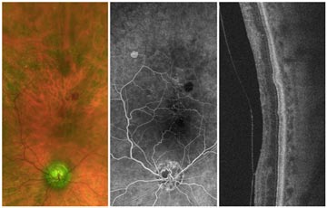
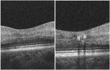
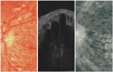
Case 24
Central Retinal Vein Occlusion (CRVO) with Neovascular Glaucoma (NVG)
Case 27
Juxtafoveal Telangiectasis (JFT)
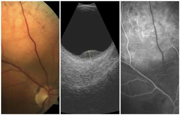
Case 28
Central Retinal Artery Occlusion (CRAO)
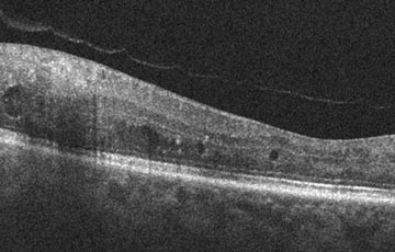
Case 29
Non-proliferative Diabetic Retinopathy (NPDR) + Retinal Vein Occlusion (RVO)
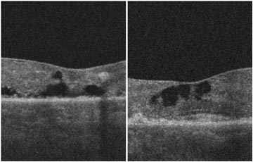
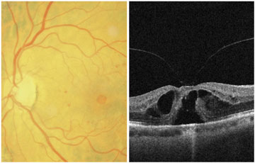
Case 30
Epiretinal Membrane with Schisis
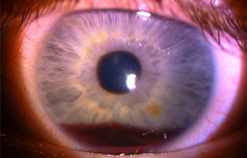
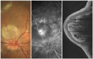
Case 31
Neovascularization of the Iris (NVI)
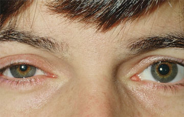
Case 32
Horner's Syndrome
Case 33
Central Retinal Vein Occlusion (CRVO)
Case 34
Choroidal Neovascularization (CNV)
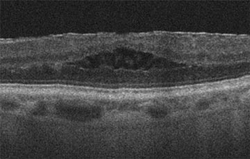
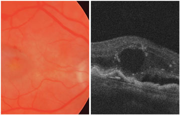
Case 35
Macular Hole